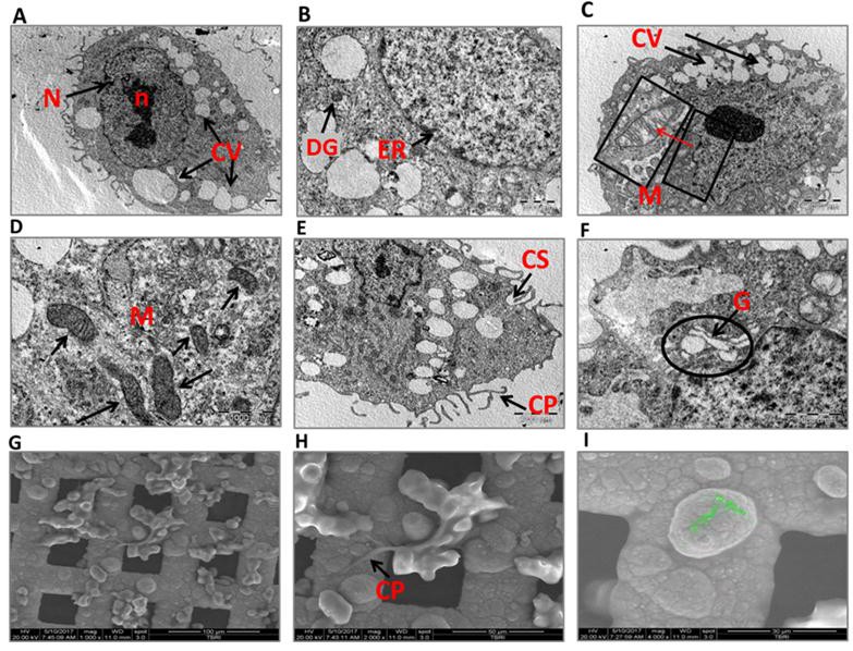
Fig. 4. Electron micrographs of pericytes showing intracellular organelles. A. Nucleus (N), nucleolus (n), cytoplasmic vesicles (CV). B. magnified image showing the endoplasmic reticulum (ER) and dense granules (DG). C and D. Mitochondria (M) are located close to the nucleus; the red arrow shows a magnified mitochondrion with its transverse cristae. E. The cytoplasmic processes (CP) of pericytes form the caveolae system (CS). F. Golgi bodies (G) are located close to the nucleus. G-I. Images showing the pericyte topography. H. Magnification of the image shown in G, (CP), cytoplasmic processes. I. Image showing the pericyte diameter, X4000.